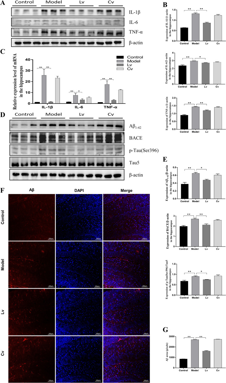
Fig. 7.
Lateral ventricle administration of BMSC-exos reduced the expression levels of IL-1β, IL-6 and TNF-α, Aβ, and p-Tau in the hippocampus of mice injected with STZ. A Typical Western Blot photos of IL-1β, IL-6 and TNF-α; B Quantitative analysis of the protein expression of IL-1β, IL-6 and TNF-α. C Quantitative analysis of the mRNA expression of IL-1β, IL-6 and TNF-α. D Typical Western blot photographs of Aβ1-42, BACE and p-Tau (ser396). E Quantitative analysis of the protein expression of Aβ1-42, BACE, and p-Tau (ser396) expression level. F Fluorescence detection of Aβ in hippocampus. G Quantification of the pixels of Aβ positive area. Data are presented as means ± SEM, with n = 3 in each group (*P < 0.05, **P < 0.01)

