Figure 6.
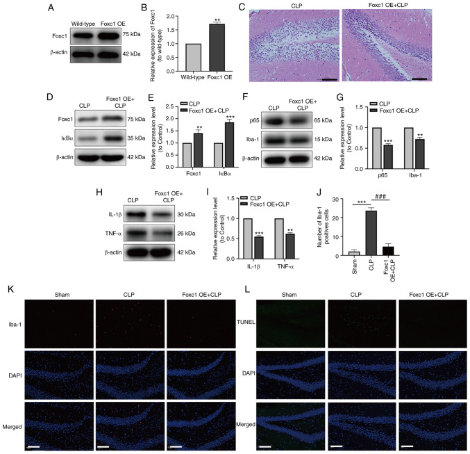
Overexpression of Foxc1 inhibits microglial migration, suppresses the inflammatory response and neuronal apoptosis in the hippocampus of mice with sepsis-associated encephalopathy through the NF-κB pathway. The expression of Foxc1 in the hippocampus of mice with or without Foxc1 overexpression was analyzed by (A) western blot analysis and (B) was semi-quantified, **P<0.01 vs. wild-type group. (C) H&E staining of the mouse hippocampal tissue. Scale bar, 100 µm. (D) The expression of Foxc1 and IκBα in hippocampal tissue of mice was measured by western blotting (E) and quantified. (F) The expression of p65 and Iba-1 in the hippocampal tissues of the mice was measured by western blotting (G) and quantified. (H) The expression of IL-1β and TNF-α in the hippocampal tissues of mice was measured by western blot analysis (I) and quantified. (K) Immunofluorescence staining quantification of Iba-1 in the mouse hippocampal tissue. (J) Representative immunofluorescence images of Iba-1 staining, where red dots represent microglia. Scale bar, 20 µm; n=3/group. (L) TUNEL staining, where green dots represent apoptotic neurons. Scale bar, 20 µm. **P<0.01 and ***P<0.001 vs. CLP or as indicated; ###P<0.001 as indicated. Foxc1, Forkhead box C1; OE, overexpression; IκBα, NF-κB inhibitor α; CLP, cecal ligation and perforation; Iba-1, allograft inflammatory factor 1.

