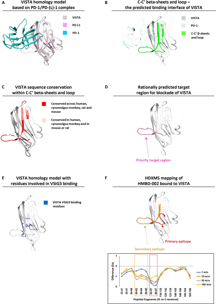
Figure 2.
HMBD-002 is a unique anti-VISTA antibody, immunoengineered to bind to a rationally predicted functional epitope that is species-conserved and overlaps with key residues for VISTA interaction with VSIG3. (A) VISTA homology model (gray) superimposed with the complex of PD-L1 (purple) bound to its ligand PD-1 (cyan) from PDB:5IUS. (B) Front C-C’ β sheets and loop of VISTA protein constituting the predicted binding interface for physiologically relevant binding partners (highlighted in green). (C) Three-dimensional (3D) overlay model of VISTA protein from mouse, rats, cynomolgus monkey and humans showing sequence conservation of the C-C’ β sheets and loop. (D) 3D model of the computationally predicted target region for antibody blockade of VISTA. (E) Predicted VISTA homology model with residues implicated in VSIG3 binding. (F) Epitope mapping analysis of HMBD-002 bound to VISTA using hydrogen–deuterium exchange mass spectrometry. PD-1, programmed cell death protein-1; PD-L1, programmed death-ligand 1; VISTA, V-domain Ig suppressor of T cell activation; VSIG3, V-Set and Immunoglobulin domain containing 3.

