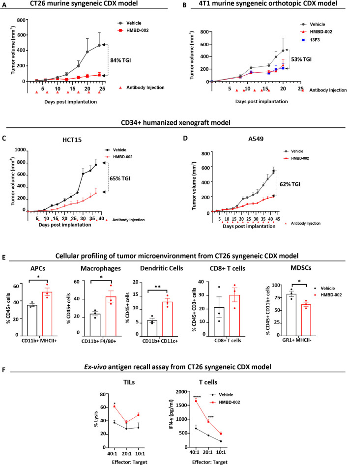
Figure 6.
HMBD-002 demonstrates strong antitumor responses as a single agent in multiple CDX models of solid tumors and remodels the TME to an antitumorigenic, proinflammatory phenotype. (A–D) CDX models used in the study included female BALB/c mice, subcutaneously implanted with CT26 (A); Female Balb/c mice orthotopically implanted with VISTA O/E 4T1 cells (B) and CD34 engrafted humanized female HiMice, subcutaneously implanted with either A549 (C) or HCT15 (D) cells. Mice were randomized and dosed with concentrations of test articles at indicated time points. Tumor volumes were measured two times a week. Each data point represents the mean tumor volume±SEM from n=10 mice. (E) Tumor-infiltrating leukocyte (TIL) populations in the TME of CT26 syngenic CDX model profiled by FACS. Data shown are mean of n=3 and error bars are SEM. (F) CT26 Antigen recall assay measured by ex vivo culture of TILs (CD45+) with CT26 cells to measure lysis or culture of tumor enriched T cells (CD4+/CD8+) with CT26 cells to measure T cell activation via IFN-γ secretion using ELISA. Data shown are mean of n=3 and error bars are SEM. All p values were obtained by unpaired t-tests, *p<0.05, **p<0.01. Each data point represents a mouse. CDX, cell-derived xenograft; IFN, interferon; MDSCs, myeloid-derived suppressor cells; TME, tumor microenvironment; VISTA, V-domain Ig suppressor of T cell activation.

