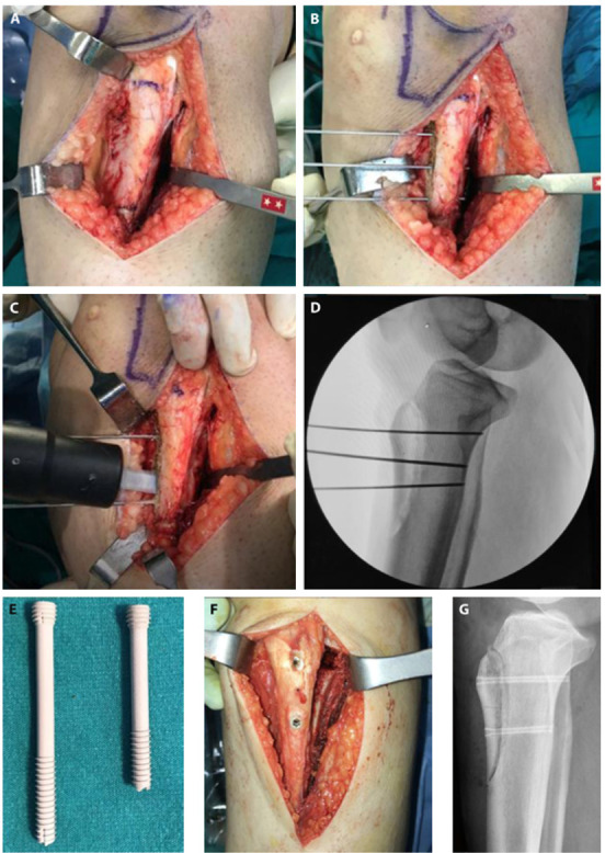
Figure 1.

(a) Identification and marking of tibial tubercle (b) Insertion of K wires. (c) Creation of osteotomy (d) Fluoroscopic control (e) Appearance of 4.8 mm bioabsorbable magnesium compression screws (f) Fixation of the TTO and postoperative lateral radiograph (g).
