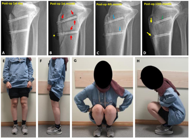
Figure 2.

(a) Immediate postoperative radiograph. (b) First month follow-up showing radiolucent zone around the screws (red arrows) and gas accumulation within the subcutaneous tissue (yellow asterix). (c) Fourth month follow-up. Decreased radiolucency (blue arrows) and union of the osteotomy was seen. (d) Final follow-up demonstrated consolidation of the osteotomy, absorption of the screw heads and radiolucent zone around the screw (green arrow). (e-h) Final clinical appearance of the patient.
