
Figure 4.
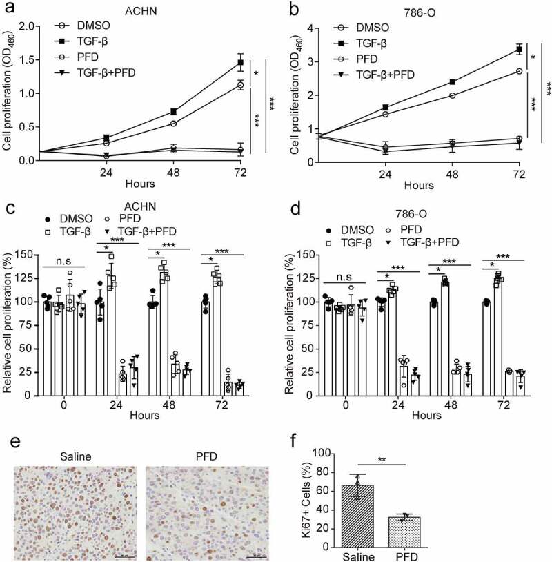
PFD inhibits proliferation of renal cancer cells.
(A) and (B) The proliferation of renal cancer cells was performed by CCK-8 assays in 786-O and ACHN cells treated with PFD in the absence or presence of TGF-β for different times (0, 24, 48, 72 h). (C) and (D) The quantification of relative cell proliferation. (E) Representative pictures of Ki67 immunohistochemical staining in paraffin-embedded tumor tissues. (F) Quantification of the percentage of Ki67-positive cells within tumor tissues. Data represents the means ± SD. *P < .05; **P < .01; ***P < .001.