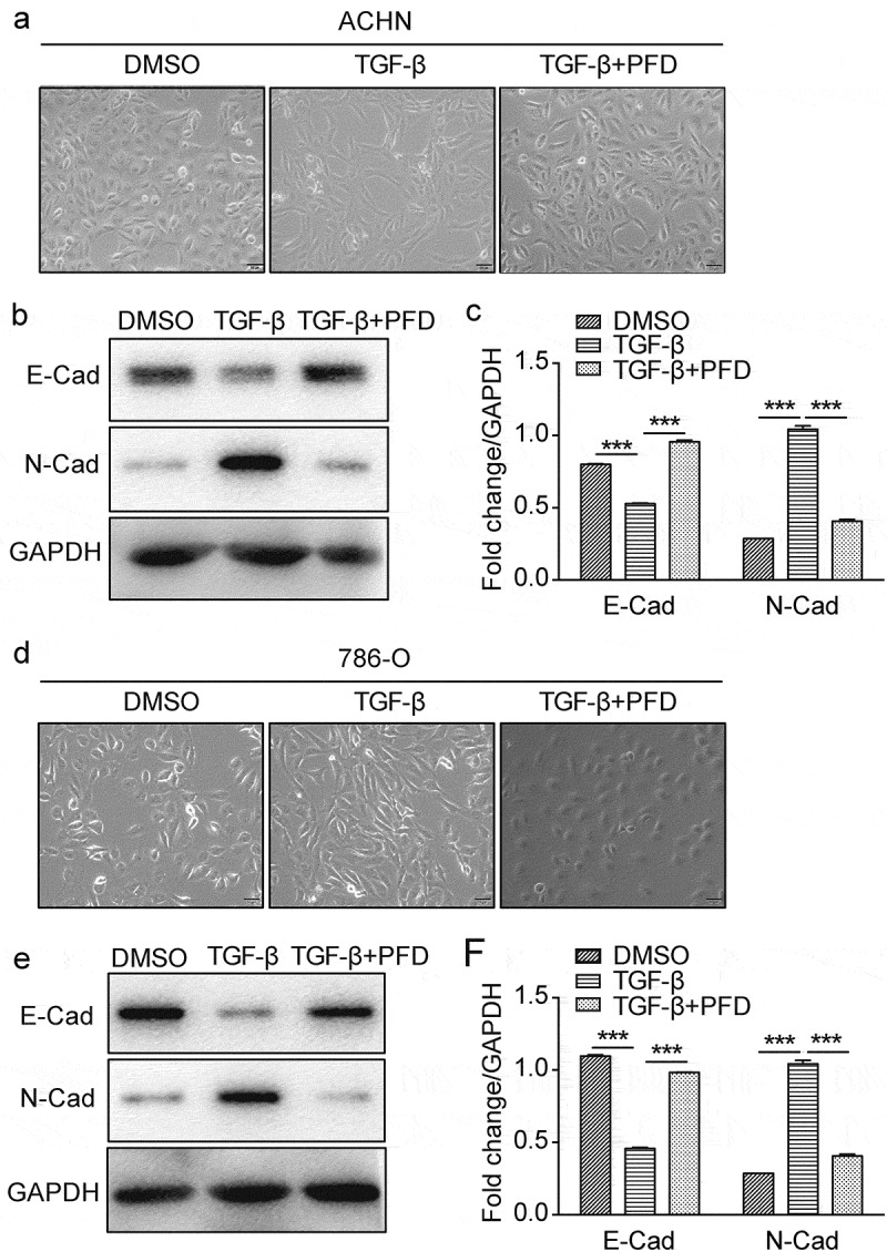
Figure 6.

PFD suppresses TGF-β-induced EMT in renal cancer cells.
(A) and (D) The morphological change of the EMT model of renal cancer cells induced by TGF-β was observed under the phase-contrast microscope after treated with PFD. (B/C) and (E/F) PFD suppresses TGF-β-induced EMT on ACHN and 786-O. Western blot analysis for the protein expression of indicated EMT-related markers (E-cadherin, N-cadherin) in 786-O and ACHN cells. Data represents the means ± SD. ***P < .001.
