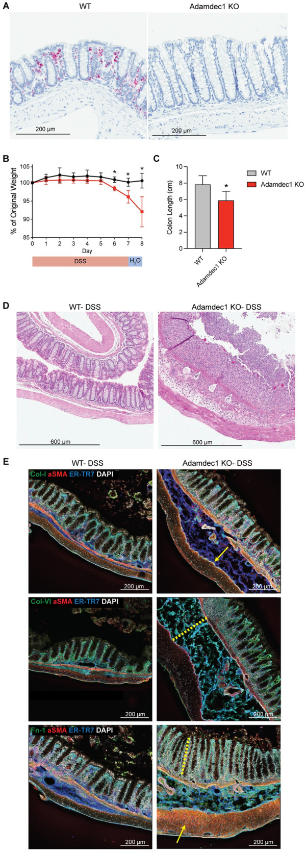
Fig 6. Adamdec1 is required for matrix remodeling and healing in response to epithelial injury.

(A) ISH of Adamdec1 in WT or Adamdec1 KO colon at basal state (water-fed mice). Scale bar, 200 μm. n = 2 per cohort. (B) Weight loss in Adamdec1 KO versus WT mice administered 2% DSS for 7 days and followed by H2O for 7 days. n = 4 per cohort. Mann–Whitney test, *p < 0.05. (C) Colon length in Adamdec1 KO versus WT mice administered DSS as described above. n = 8 per cohort. Mann–Whitney test, *p < 0.05. For source data for panels B and C, see S1 Data. (D) HE staining of representative images of colon following DSS in WT and Adamdec1 KO mice. Mice were administered 2% DSS for 7 days, H2O for 1 day, and killed on day 8. Scale bar, 600 μm. n = 4 per cohort. (E) IF staining was performed on colons from Adamdec1 KO and WT mice with indicated markers. Mice were administered 2% DSS for 7 days and killed on day 7. αSMA (red), ER-TR7 (blue), DAPI (gray). Indicated ECM component (green). (Top row) Collagen type I (green). Yellow arrow denotes submucosal ECM accumulation. (Middle row) Collagen type VI (green). Yellow dotted line denotes submucosal thickening and edema. (Bottom row) Fibronectin (green). Yellow dotted line denotes hyperplastic response. Yellow arrow denotes muscle thickening. Scale bar, 200 μm. n = 3 per cohort, representative of 2 experiments. DSS, dextran sulfate sodium; ECM, extracellular matrix; HE, hematoxylin–eosin; IF, immunofluorescence; ISH, in situ hybridization; KO, knockout; WT, wild-type.
