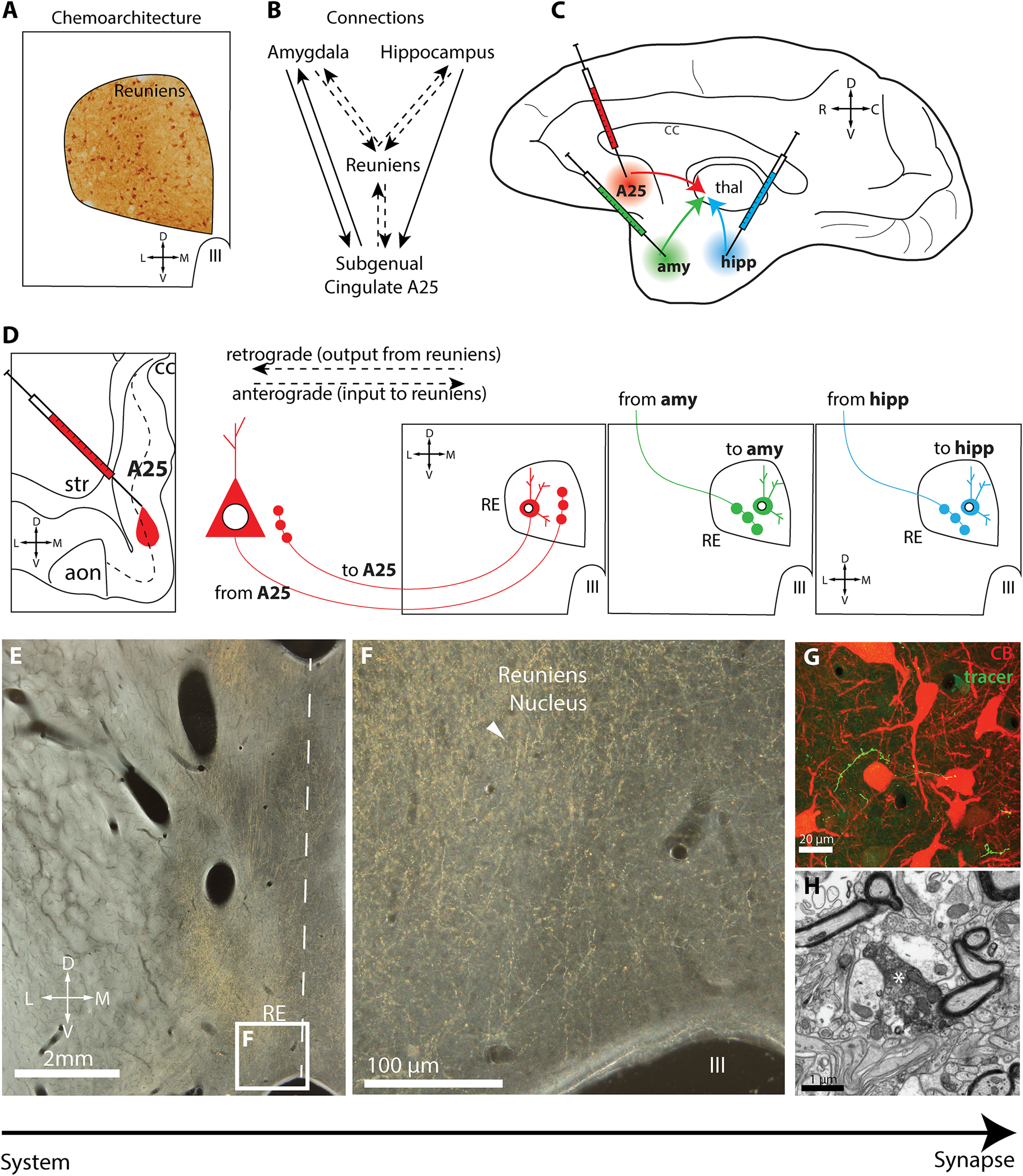
Figure 1.

Experimental design. A, Schematic depicts immunohistochemical labeling in the reuniens nucleus to characterize its chemoarchitecture for CB, CR, PV, and GABA. B, Circuit organization schematic linking amygdala, hippocampus, and subgenual cingulate A25 with RE. Dashed lines indicate pathways under investigation in this study. C, Schematic shows injection sites of neural tracers in A25, amygdala, and hippocampus. D, Schematic depicts retrograde and anterograde transport from injection sites to RE. E, Low-power photomicrograph taken using darkfield microscopy of tracer-labeled axonal terminations (yellow gold) in RE for pathway mapping and high-resolution ultrastructural investigation. Dashed line depicts midline. F, Inset, From E, tracer-labeled axonal terminations (yellow gold) in RE (white arrowhead). G, Photomicrograph taken using confocal microscopy shows tracer-labeled axon terminations (green) comingling with matrix CB neurons (red) in RE. E–G, Our strategy, which uses methods with increasing resolution to study the properties of these pathways, from system to synapse. H, Photomicrograph taken using EM shows tracer-labeled axon termination (white asterisk) in RE. aon, Anterior olfactory nucleus; amy, amygdala; C, caudal; cc, corpus callosum; D, dorsal; hipp, hippocampus; III, third ventricle; L, lateral; M, medial; R, rostral; str, striatum; thal, thalamus; V, ventral.
