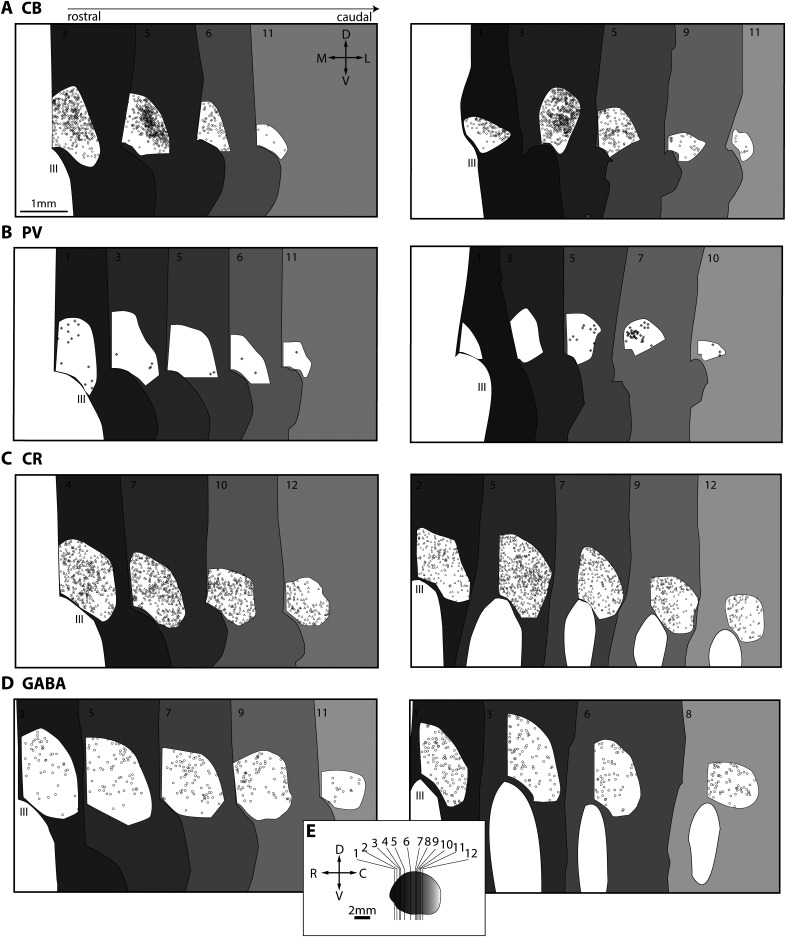
Figure 4.
Maps of the PV, CB, CR, and GABA neurons in reuniens nucleus. Coronal sections through RE are depicted from rostral (dark gray) to caudal (light gray). A, CB neurons in RE in two cases (left, AZ; right, BD). Compass and scale apply in A–D. B, PV neurons RE in two cases (left, AZ; right, BD). C, CR neurons in RE in two cases (left, BV; right, BW). D, GABA neurons in RE in two cases (left, BV; right, BW). E, Planes of cut for plots in A–D. C, Caudal; D, dorsal; III, third ventricle; L, lateral; M, medial; R, rostral; V, ventral.

