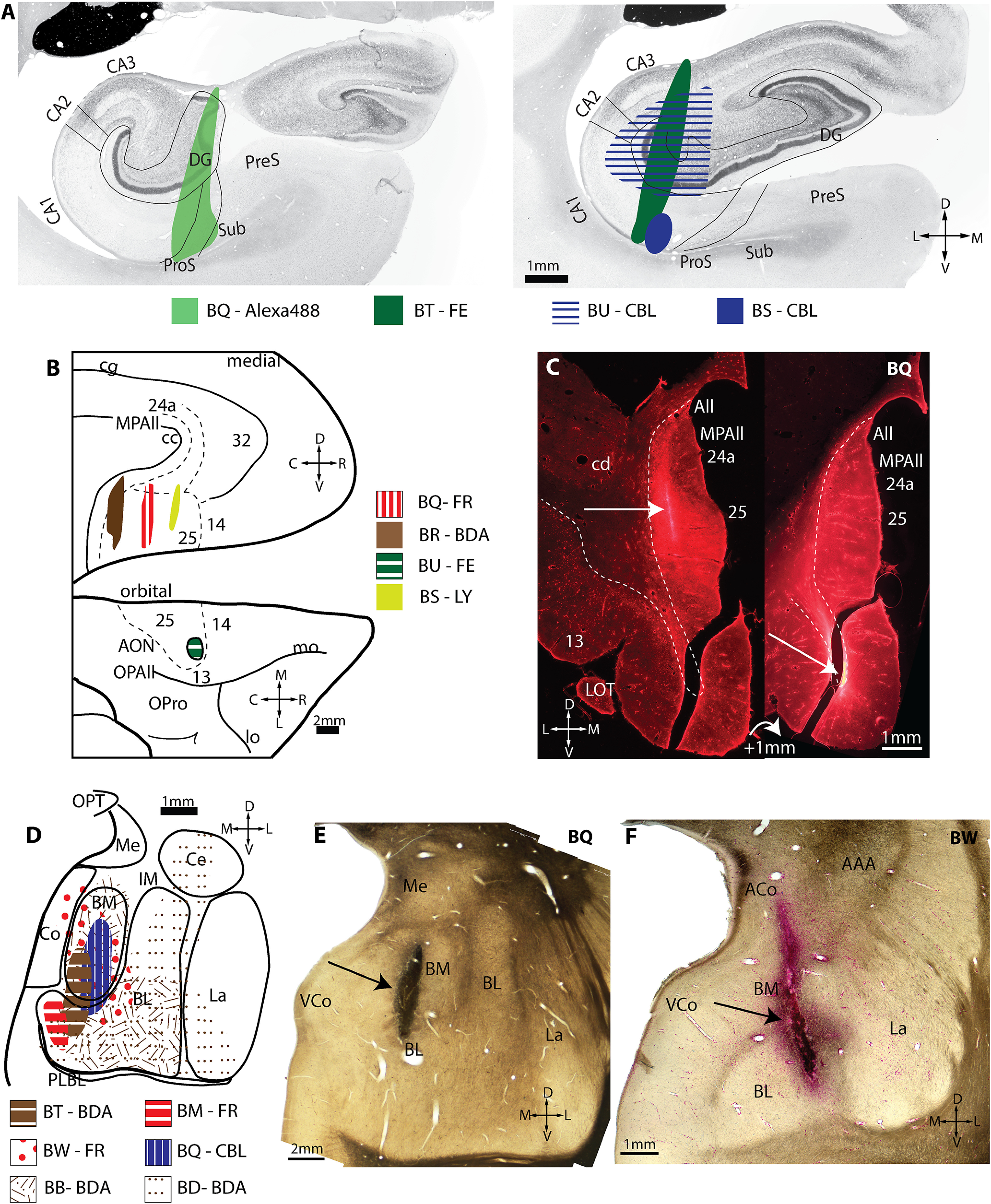
Figure 5.

Injection sites in hippocampus, A25, and amygdala. A, Injection sites in two planes through the hippocampus. B, Injection sites in normalized A25 space. Top, Medial surface of the rhesus monkey brain. Bottom, Orbital surface. C, Fluorescent photomicrographs of two coronal sections through A25 depict the extent of the injection site in medial A25. Dashed line delineates the boundary with the white matter. White arrow indicates injection site. D, Injection sites in normalized amygdala space. E–F, Brightfield photographs of wet-mounted coronal sections through the amygdala depict injection sites (black arrows). AAA, Anterior amygdaloid area; ACo, anterior cortical nucleus of amygdala; All, Allocortex; AON, anterior olfactory nucleus; BL, basolateral nucleus of the amygdala; BM, basomedial nucleus of the amygdala; C, caudal; CA, cornu ammonis; CBL, Cascade Blue; cc, corpus callosum; cd, caudate; Ce, central nucleus of the amygdala; cg, cingulate sulcus; Co, cortical nucleus of the amygdala; D, dorsal; DG, dentate gyrus; FE, Fluoroemerald; FR, Fluororuby; IM, intercalated masses of the amygdala; L, lateral; La, lateral nucleus of the amygdala; lo, lateral orbital sulcus; LOT, lateral olfactory tract; LY, Lucifer yellow; M, medial; Me, medial nucleus of the amygdala; mo, medial orbital sulcus; MPAll, medial periallocortex; OPAll, orbital periallocortex; OPro, orbital proisocortex; OPT, optic tract; P, posterior; PLBL, paralaminar basolateral nucleus of the amygdala; ProS, prosubiculum; PreS, presubiculum; R, rostral; Sub, subiculum; uCA, uncal cornu ammonis; V, ventral; VCo, ventral cortical nucleus of the amygdala.
