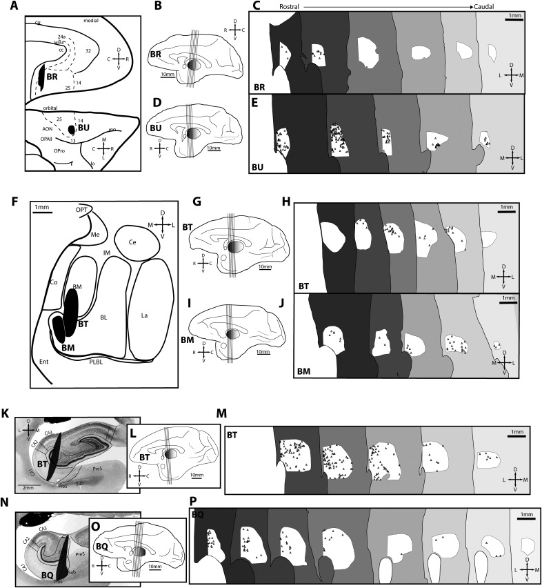
Figure 7.
Maps of projection neurons in reuniens nucleus directed to A25, amygdala, and hippocampus. A, Normalized A25 space depicts location of injection sites (black) in A25 that were used to map retrogradely labeled neurons in RE. BR featured a posterior medial injection site, whereas BU featured an anterior orbital injection site. Dashed lines depict areal boundaries. B, Medial surface (case BR) with lines depicting planes of cut for sections sampled in C. C, Retrogradely labeled neurons in RE directed to medial A25 shown from rostral (left, dark gray) to caudal (right, light gray) levels (case BR). D–E, Organization as above, but for tracer injection in orbital A25 (case BU). F, Normalized amygdala space depicts location of injection sites (black) used to map retrogradely labeled neurons. One case (BT) featured an injection in the basomedial and ventral medial sector of the basolateral nucleus. The other case (BM) featured an injection in the ventral medial sector of the basolateral nucleus. G–H, As above, but for case BT. I–J, As above, but for another amygdalar case (case BM). K, Injection site (black) in the hippocampus of case BT used to map retrogradely labeled neurons after tracer injection in CA3, DG, and CA1. L–M, As above, but for hippocampal case BQ. N, Injection site (black, case BQ) in used to map retrogradely labeled neurons in RE directed to hippocampus after tracer injection in CA1, DG, prosubiculum, and subiculum. O–P, As above, but for case BQ with hippocampal injection as shown in N. AON, Anterior olfactory nucleus; BL, basolateral nucleus of the amygdala; BM, basomedial nucleus of the amygdala; C, caudal; CA, cornu ammonis; Ce, central nucleus of the amygdala; cg, cingulate sulcus; Co, cortical nucleus of the amygdala; D, dorsal; DG, dentate gyrus; Ent, entorhinal cortex; III, third ventricle; IM, intercalated masses of the amygdala; L, lateral; La, lateral nucleus of the amygdala; lo, lateral orbital sulcus; M, medial; Me, medial nucleus of the amygdala; mo, medial orbital sulcus; MPAll, medial periallocortex; OPAll, orbital periallocortex; OPro, orbital proisocortex; OPT, optic tract; PLBL, paralaminar basolateral nucleus of the amygdala; ProS, prosubiculum; PreS, presubiculum; R, rostral; Sub, subiculum; V, ventral.

