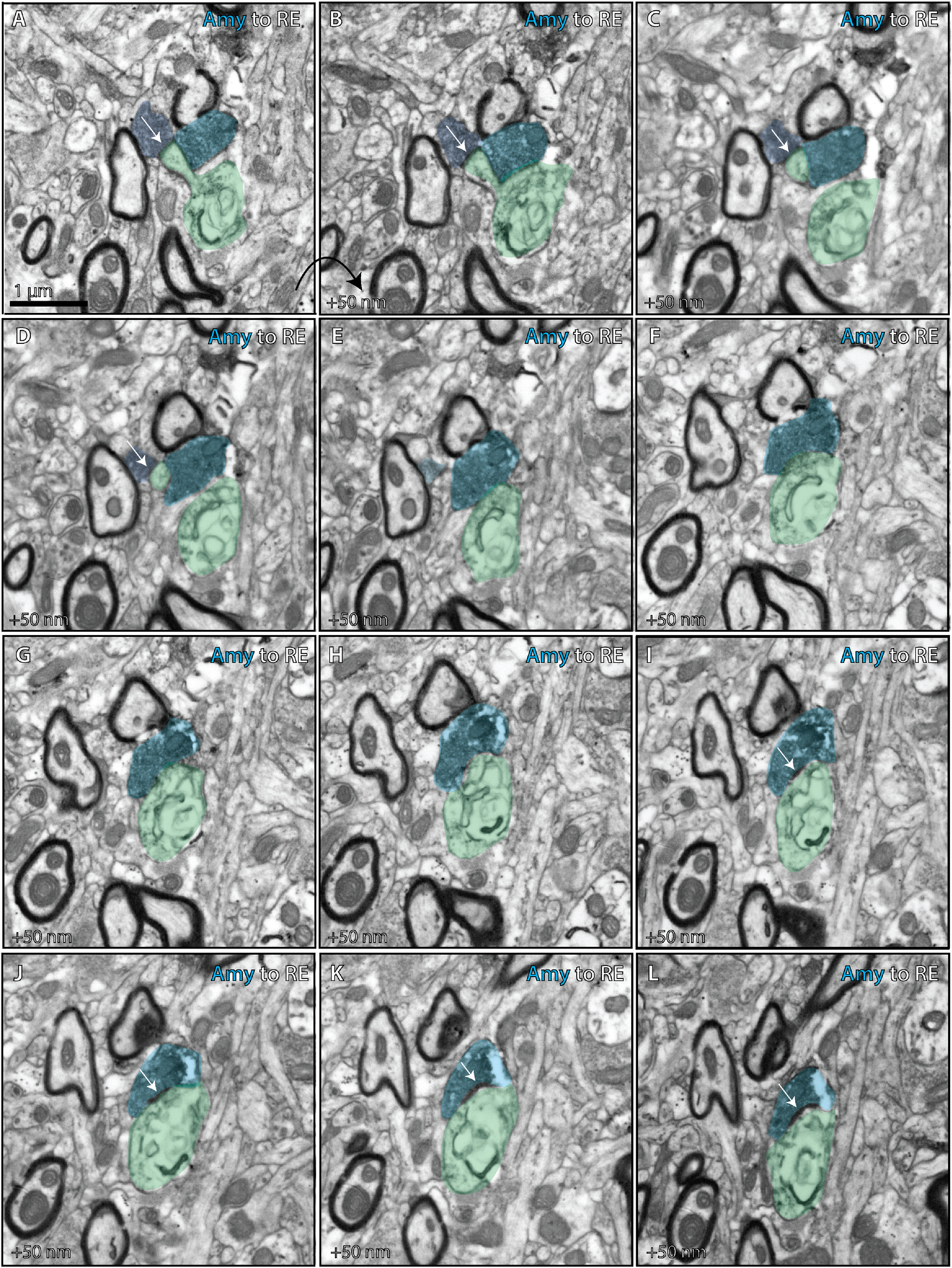
Figure 9.

Ultrastructural analysis of amygdalar axon in the nucleus reuniens. Photomicrographs from serial EM depict ultrathin (50 nm) sections through the RE (case BQ, CBL tracer). Together they depict an amygdalar axon forming distinct terminations on a thorn and shaft of the same dendrite in RE. A–D, One termination (dark blue) from the amygdalar axon forms an asymmetric synapse (white arrows) on a thorn of an RE dendrite (green). E–L, A distinct termination from the same amygdalar axon (lighter blue) forms an asymmetric synapse (white arrows) on the shaft of the RE dendrite (green). Amy, Amygdala; CBL, Cascade Blue
