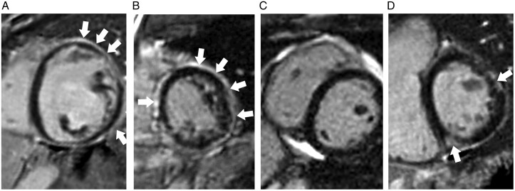
Figure 2.
Examples of desmoplakin cardiomyopathy on cardiac magnetic resonance. (A) Left dominant: left ventricle is dilated and dysfunctional (LVEF = 25% and LVEDVi = 188 mL/m2. The RV is normal. Extensive subendocardial LGE in the left ventricle at mid cavity. (B) Biventricular involvement: both left and right ventricular function was reduced (LVEF 49% and RVEF 43%). Extensive subepicardial and mid-wall LGE in a non-ischaemic pattern throughout the entire circumference of the left ventricle at mid-cavity. (C) Right dominant: left ventricle is normal but RVEF is reduced (34%) although RVEDVi remains normal (79 mL/m2) No evidence of LGE. (D) Normal ventricular function but LGE present.

