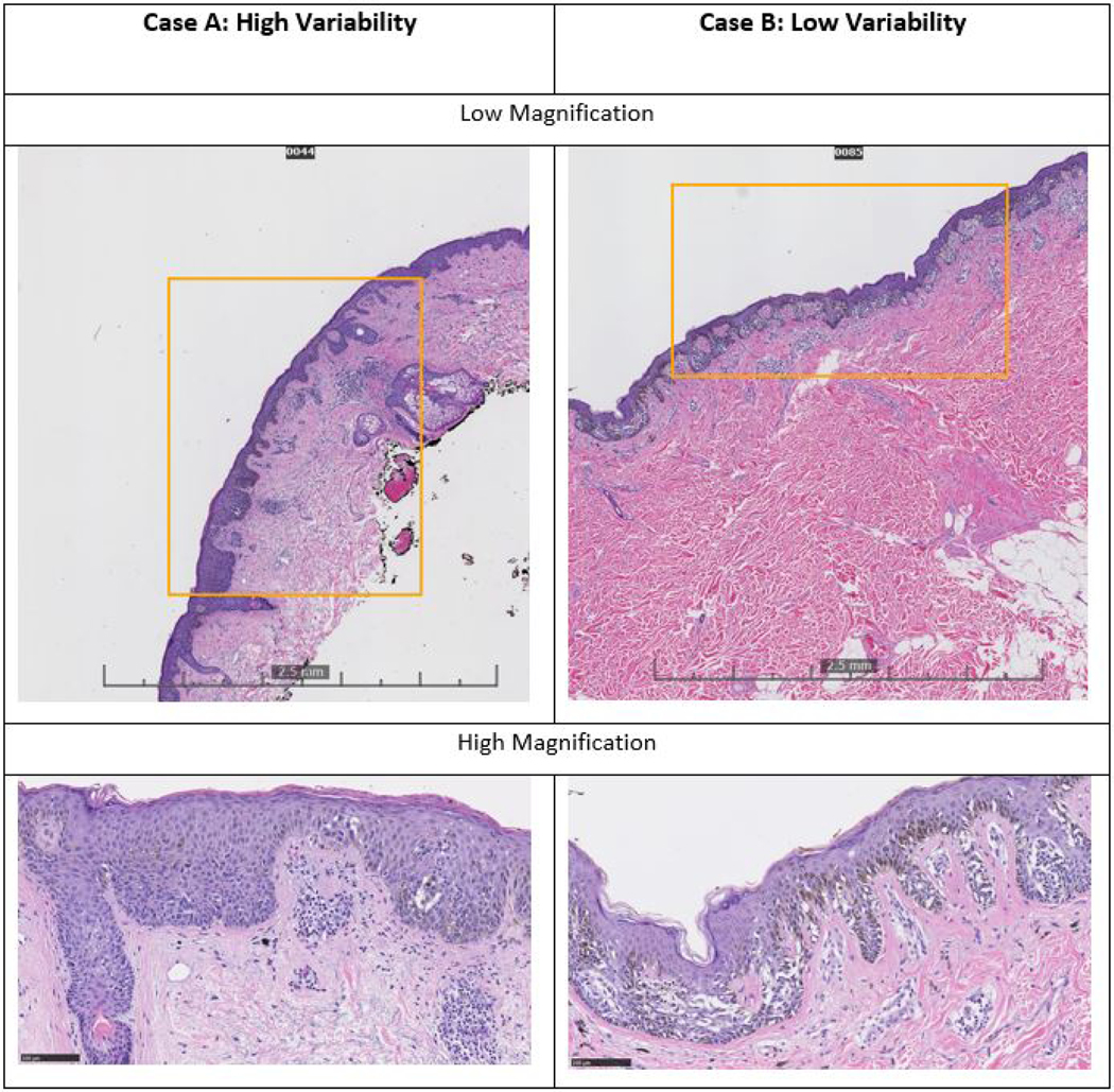
Figure 2: Two Examples of Melanoma In Situ Glass Slide Biopsies evoking High and Low Variability in Treatment Suggestions by the Study Participants.

Both cases were diagnosed as melanoma in situ by consensus panel and 17 out of 18 pathologists. For Case A, 29% of participating pathologists suggested a lower treatment (<0.5cm) and 18% suggested a higher treatment (≥ 1cm margin) than NCCN guidelines, showing relatively large variability in treatment suggestions. Case A is relatively small (~4mm) and does not have an obviously atypical melanocytic proliferation at low magnification. Higher magnification allows the identification of a poorly circumscribed intraepidermal melanocytic proliferation. In contrast, Case B had a smaller proportion of pathologists (12%) rendering a treatment suggestion lower than NCCN guidelines, and no pathologists suggested a higher treatment than NCCN guidelines. Case B is a larger lesion (~8mm) that more readily is identifiable as an atypical lentiginous and nested proliferation with pagetoid scatter.
