Figure 2.

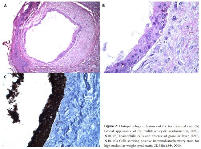
Histopathological features of the trichilemmal cyst. (A) Global appearance of the multilayer cystic neoformation, H&E, ×10. (B) Eosinophilic cells and absence of granular layer, H&E, ×40. (C) Cells showing positive immunohistochemistry stain for high molecular weight cytokeratin CK34Be12+, ×40.
