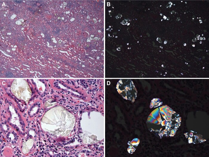
FIGURE 1:
Renal oxalosis: (A) Massive deposition of oxalate crystals is noted in tubules with associated advanced chronic tubulointerstitial disease with atrophy and dropout of tubules and prominent interstitial fibrosis and nonspecific inflammation (H&E, bright field, 40X). (B) Same area visualized under polarized light reveals numerous intratubular crystals (H&E, polarized light, 40X). (C) Intratubular oxalate crystals are often transparent or reveal yellow or gray color, with needle or other shapes of crystals (H&E, bright field, 600X). (D) Same area visualized under polarized light reveals colorful crystals (H&E, polarized light, 600X)

