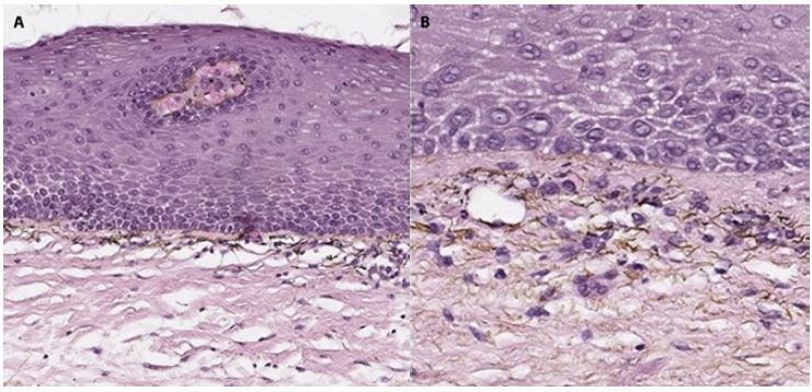
Figure 2.

Penile exogenous pigmentation: histopathological findings consisting of epithelium hyperplasia, sclerosis and brownish-black pigment in the superficial chorion, with no evidence of atypical melanocytic proliferation. (A) Hematoxylin and eosin stain original magnification ×17. (B) Hematoxylin and eosin stain original magnification ×32.
