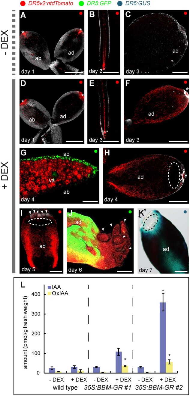
Figure 4.

BBM expression enhances DR5 auxin response and IAA biosynthesis. Confocal laser scanning micrographs of cotyledons or roots from 35S:BBM-GR DR5 seedlings grown without (A–C) and with (D–J) DEX. D–F, H and I are images of DR5v2:ntdTomato cotyledons or roots. G and J, These are images of DR5:GFP cotyledons. The images in (G) and (J) are counterstained with FM4-64. K, Light image of DR5:GUS expression in the cotyledon of a DEX-treated 35S:BBM-GR seedling. Samples were counter stained with SR2200 (gray, A–F, H and I) or outlined using red autofluorescence (G and J). The dashed ellipses in (H), (I), and (K) indicate the DR5 minimum. Small embryogenic protrusions are indicated with arrowheads in (I) and (J). va, vascular tissue; asterisks autofluorescence. Scale bars, 200 μm. L, IAA and oxIAA concentrations in seedlings of WT Col-0 and two 35S:BBM-GR lines grown in the absence or presence of DEX (three technical replicates, each 200 mg). *, samples that showed statistically significant differences in IAA or oxIAA concentrations compared to the non-DEX treated 35S:BBM-GR control (Student’s t test, p < 0.05). Error bars represent the standard deviation of the replicates.
