Figure 7.
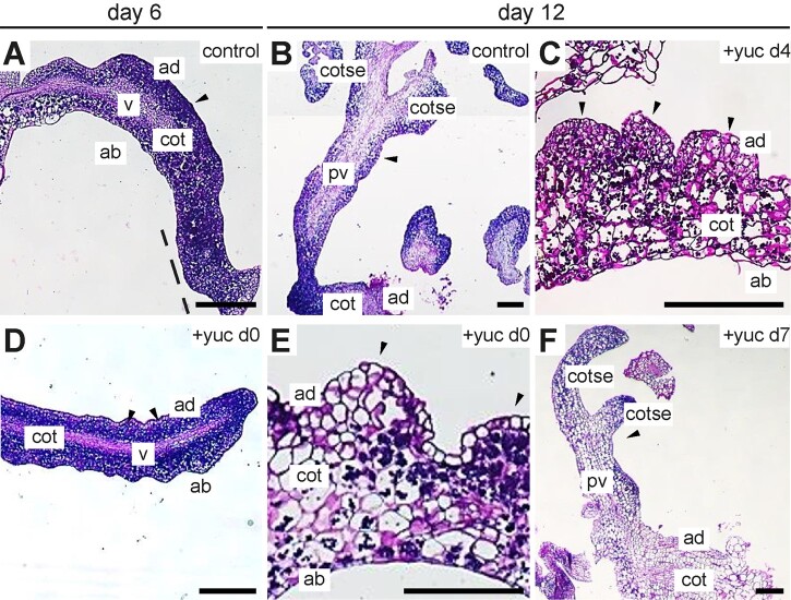
YUC-dependent auxin biosynthesis is required for the formation of histodifferentiated somatic embryos. Light micrographs of thin cross sections of the cotyledons of DEX and YUC inhibitor (yucasin)-treated 35S:BBM-GR explants fixed on the days indicated above the images. The day of culture and the yucasin treatment (100 µM) is shown above and in the image panels, respectively. A and B, Explants from control samples treated with DEX from Day 0 until the end of the culture on Day 14. Panel B is a composite of different images from the same section. C–F, Explants from samples treated with DEX from Day 0 to Day 14, to which YUC enzyme inhibitor was added on Day 0 (D and E), Day 4 (C), or Day 7 (F). Black arrowhead, growth protrusions (A, C–E) and somatic embryos (B and F); cot, cotyledon; cotse, cotyledons of somatic embryos; v, vascular (A and D); pv, provascular tissue (B and F); dotted line, proliferating cotyledon tip. Scale bars, 200 μm.

