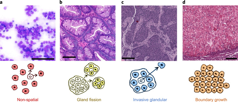
Fig. 1. Representative regions of histology slides from human tumours exemplifying four different kinds of tissue structure and manners of cell dispersal.
a, Acute myeloid leukaemia, M2 subtype, bone marrow smear. b, Colorectal adenoma. c, Breast cancer (patient TCGA-49-AARR, slide 01Z-00-DX1). d, Hepatocellular carcinoma (patient TCGA-CC-5258, slide 01Z-00-DX1). Image a is courtesy of Cleo-Aron Weis; image b is copyright St Hill et al. (2009)91 and is used here under the terms of a Creative Commons Attribution License; images c and d were retrieved from TCGA at https://portal.gdc.cancer.gov, with brightness and contrast adjusted linearly for better visibility. Scale bars, 100 μm. The illustration below each histology image describes the corresponding types of spatial structure and cell dispersal.

