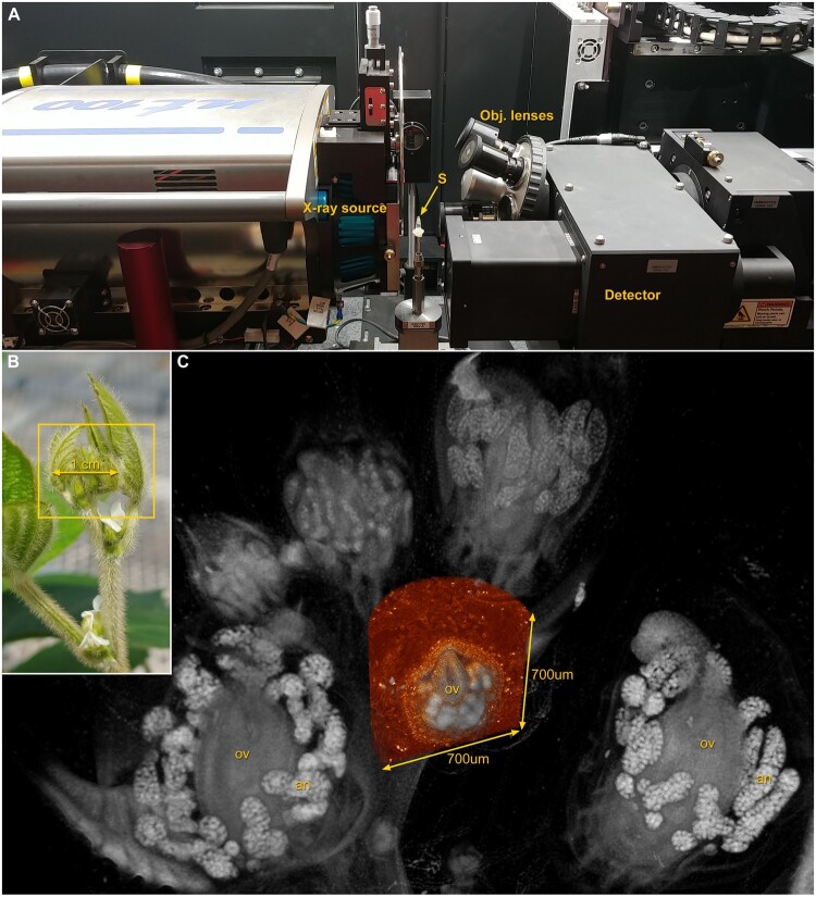
Figure 1.
X-ray microscope (XRM) and multiscale imaging of soybean (Glycine max) for floral anatomy. A, View inside the Zeiss XRM used to image soybean samples in B and C with the positions of the sample (S), X-ray source, detector, and objective lenses indicated. B, Axillary bud complex of soybean illustrates how an entire intact sample (box) can be prepared and imaged in high resolution 3D with XRM (C), followed by a region-of-interest scan at a higher magnification (orange), without removing the sample from the instrument. Reproductive organs such as ovaries (ov) and pollen-bearing anthers (an) are readily visualized.

