Figure 1.

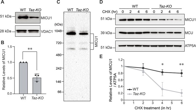
MICU1 abundance and stability are reduced in C2C12 Taz-KO cells. (A) SDS-PAGE immunoblot analysis of MICU1 in mitochondria isolated from C2C12 WT and Taz-KO myoblasts. VDAC1 is used as a loading control. (B) Quantification of relative MICU1 levels from (A) by densitometry using ImageJ software. Data shown as mean ± SD. n = 3. **P < 0.005. (C) BN-PAGE immunoblot analysis of MICU1 in digitonin-solubilized mitochondria isolated from C2C12 WT and Taz-KO myoblasts. Blot is representative of three independent experiments. (D) SDS-PAGE immunoblot analysis of MICU1 and MCU in C2C12 WT and Taz-KO myoblasts treated with 20 μg/ml cycloheximide (CHX) for the indicated time-points. ATP5A is used as loading control. (E) Quantification of relative MICU1 levels from (D) by densitometry using ImageJ software. Data are represented as mean ± SD. n = 3. *P < 0.05, **P < 0.005.
