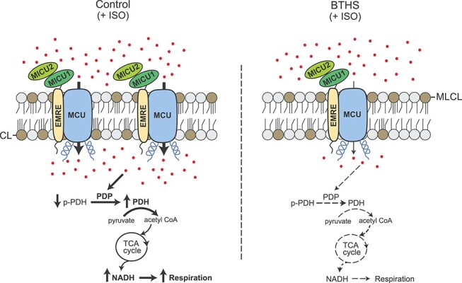
Figure 6.

A model depicting perturbed mitochondrial calcium signaling in BTHS cells. Depletion of CL and an accumulation of its precursor monolysocardiolipin (MLCL) in BTHS mitochondria result in a reduced abundance of MCU, MICU1 and MICU2. Addition of β-adrenergic agonist (ISO) fails to stimulate uptake of Ca2+ (shown as red dots) in BTHS mitochondria to the same extent as in the control mitochondria. This prevents Ca2+-dependent dephosphorylation of pyruvate dehydrogenase leading to reduced PDH activity. This in turn decreases tricarboxylic acid (TCA) cycle output in the form of NADH resulting in reduced mitochondrial respiration in BTHS mitochondria. CL, cardiolipin; MLCL, monolysocardiolipin; ISO, isoprenaline hydrochloride; PDP, pyruvate dehydrogenase phosphatase; PDH, pyruvate dehydrogenase; p-PDH, phosphorylated PDH.
