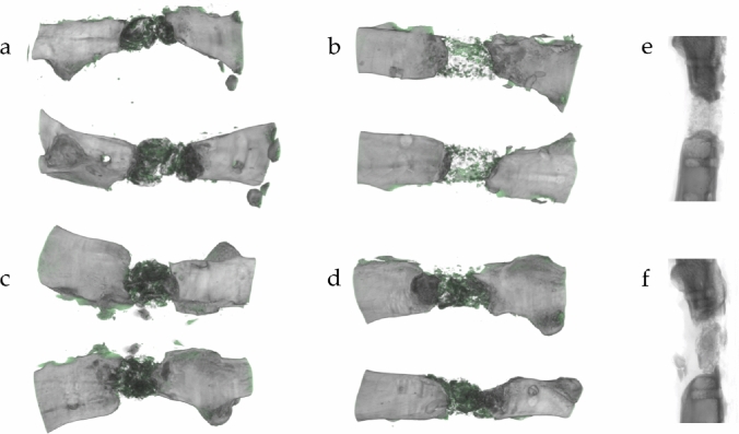
Fig. 3.
Bone formation in three-dimensional µCT of groups 1–4 (a–d). Defects treated with small granules show newly formed osseous tissue (green colored) mainly in close vicinity to the induced membrane in a tubular shape (b). Bone formation in defects with large granules is mainly concentrated within the defect (d). Two-dimensional X-ray of bones filled with small (e) and large granules (f) shows decreased contact with partially radiused blunt fracture rim and limited connection to large granules. Small granules, in contrast, create a broad contact area

