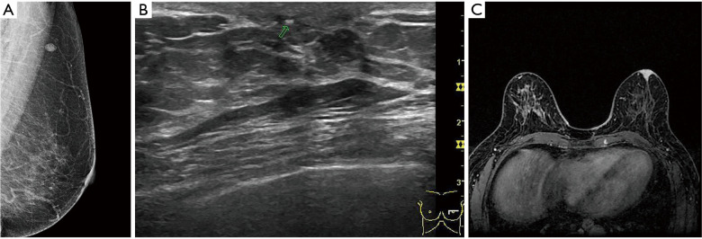
Figure 2.
The patient was a 62-year-old female with nipple erosion that had been present for 1 year. MLO of the left breast showed that the skin layer of the left nipple and areola was thickened, and the axillary lymph nodes were enlarged. (A) The ultrasound showed that the nipple was enlarged, and the strong echo light spot was behind the nipple; (B) breast magnetic resonance imaging showed abnormal enhancement of the nipple areola area; (C) Paget’s disease combined with ductal carcinoma in situ was confirmed by pathology. MLO, mediolateral oblique.

