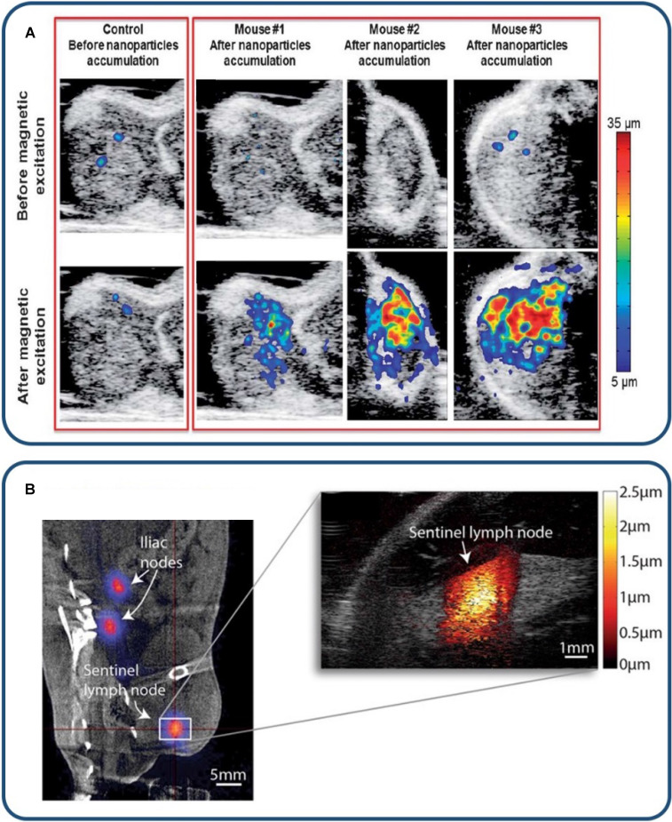
Figure 2.
MMUS/pMMUS for in vivo imaging. (A) In vivo US/pMMUS images of mice with A431 tumors. pMMUS signal detected tumors loaded with magnetic nanoparticles. Reproduced with permission from 105. Copyright 2013, Royal Society of Chemistry. (B) In vivo PET/CT image (left panel) of 68Ga-labelled SPION drainage to the sentinel lymph node. The white box indicates the magnified region in the MMUS image (right panel), where the induced magnetomotive displacement is indicated by the adjacent red color scale. Reproduced with permission from 77. Copyright 2017, Springer Nature.

