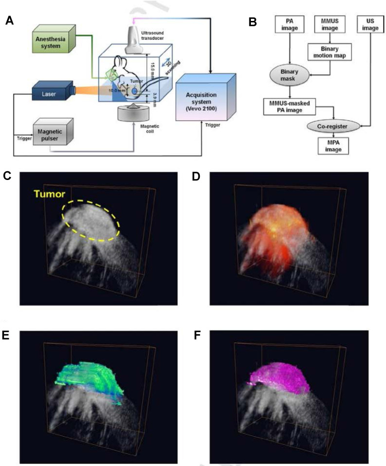
Figure 4.
Magneto-motive PA imaging (MPA) imaging of a tumor in vivo. (A) Block diagram of the in vivo MPA imaging system. (B) MPA image formation algorithm. (C) ultrasound (grayscale), (D) PA, (E) MMUS, and (D) MPA images of the tumor loaded with photomagnetic nanoparticles. Reproduced with permission from 36. Copyright 2014, Elsevier GmbH.

