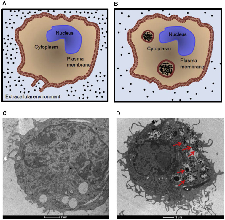
Figure 6.
Accumulation of magnetic nanoparticles within cells. (A) and (B) Diagrams depicting intracellular uptake of magnetic nanoparticles by phagocytic cells. The size of the endosomes can be as large as 5 µm. TEM images of unlabeled (C) and SPION-labeled macrophages (D). Red arrows indicate the endocytosed aggregates of SPIONs within endosomes. Reproduced with permission from 106. Copyright 2011, IOP Publishing.

