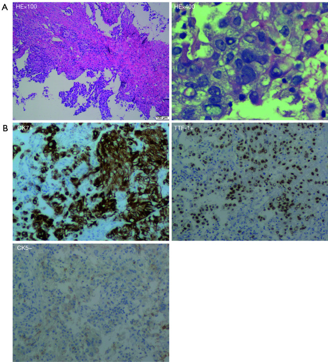
Figure 1.
Histological findings from biopsy specimens. (A) Hematoxylin and eosin (HE) staining of tumor tissue (100×, 400×) showed lung adenocarcinoma; (B) immunohistochemistry (IHC) analysis of tumor tissue showed it was positive for thyroid transcription factor-1 (TTF-1) (100×) and cytokeratin (CK)7 (100×), and negative for CK5 (100×).

