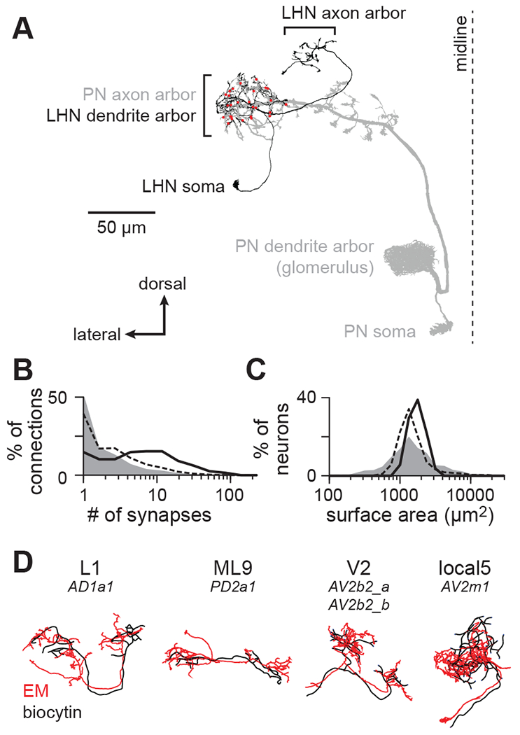
Figure 1. Matching diverse LHNs between light and electron microscopy datasets.

(A) Morphology of a single PN-LHN connection. The PN’s axon arbor targets the lateral horn, where it forms multiple synapses (red points) onto an LHN’s dendrite arbor.
(B) Distribution of the number of synapses per connection for a representative sample of connections across the hemibrain (random sample of 214,245 connections, gray), for all 17,506 PN-LHN connections (dashed line), and for the 135 PN-LHN connections matched to physiology data (solid line)
(C) Distribution of the total membrane surface area for a representative sample of neurons across the hemibrain (random sample of 1000 neurons, gray), for all 1496 LHNs (dashed line), and for the 54 LHNs matched to physiology data (black line).
(D) Example matching morphologies. Black: biocytin fill (physiology dataset). Red: EM morphology (anatomy dataset). LHN names follow a prior study25 with corresponding hemibrain names provided below24. Note that matching morphologies do not always align with hemibrain cell type boundaries. Some types (e.g., V2) correspond to multiple hemibrain types; others do not correspond to all bodyIds of a given hemibrain type.
