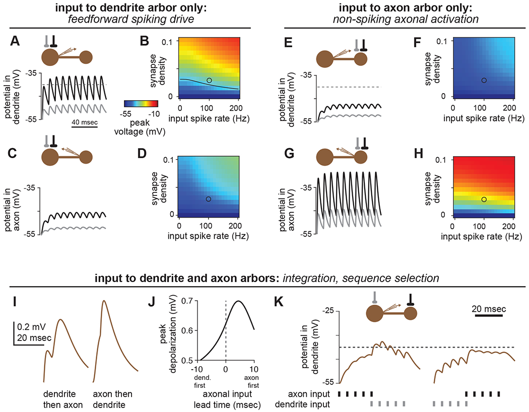
Figure 7. Predicted functions of compartmentalized neurons.

(A,B) Input to the dendrite arbor alone can readily drive spikes in local LHNs. (A) In the average local LHN, a strong connection (0.03 synapses/μm2, black trace) driven at 100Hz evokes EPSPs that depolarize the SIZ above spike threshold (−40mV). A weaker connection (0.0075 synapses/μm2, gray trace) does not reach spike threshold. (B) Peak voltages (encoded by color) obtained for dendritic stimulation for a range of synapse densities and input spike rates. Black line in the plot corresponds to the spike threshold of −40mV. The black and gray circles correspond to the black and gray traces in (A), respectively.
(C,D) As in (A-B), but with recording in the axon arbor.
(E-H) Input to the axon arbor alone cannot drive spikes, but substantially depolarizes the axon. Panels are as in (A-D), but with stimulation to the axon arbor.
(I-K) Temporal delays along the inter-arbor cable create selective responses to different input sequences. (I,J) A spike impinging on the axon before a spike impinging on the dendrite yields a larger peak depolarization at the SIZ than the opposite sequence. (K) Spike packets can depolarize the SIZ of an LHN past spike threshold in one sequence, but not the other. In each panel, the connection onto the axon is stronger than the connection onto the dendrite, to compensate for attenuation.
