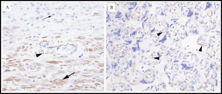
Figure 2.
Representative histologic images of vascular endothelial growth factor A (VEGF-A)–immunostained placental samples from women living with human immunodeficiency virus in Uganda. A, Placental membrane roll demonstrating positive VEGF-A staining in maternal endothelium (arrowhead) and decidua (large arrow) and negative staining in the extravillous trophoblast of the chorion laeve (small arrow) (original magnification ×40). B, Placental parenchyma demonstrating positive VEGF-A staining in chorionic villi and villous (fetal) endothelium (arrowheads) (original magnification ×40).

