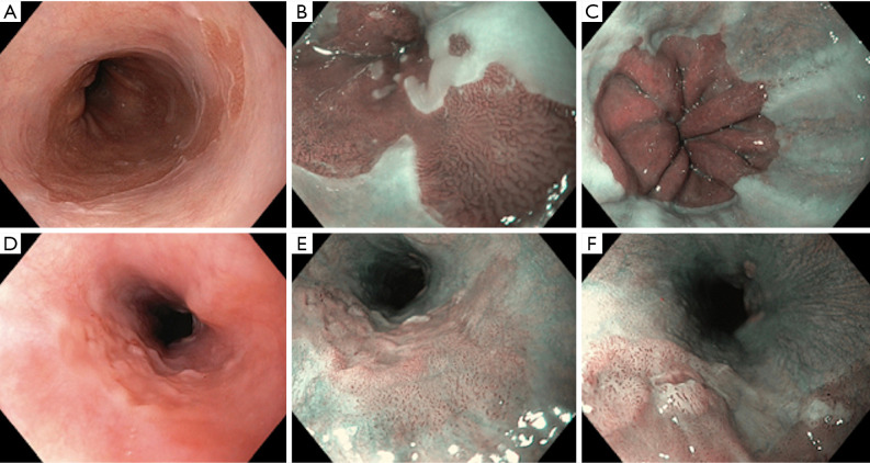
Figure 1.
Role of narrow band imaging (NBI) in esophagus (Barrett’s oesophagus, minimal change esophagitis and early oesophageal cancer). (A) Barrett’s esophagus on white light imaging (WLI); (B) Barrett oesophagus on NBI: regular ridged pit pattern with normal micro-vasculature with no evidence of dysplasia; (C) minimal change esophagitis: dilated intra-papillary capillary loop pattern (IPCLs) type II. These are enlarged but arranged in a linear regular fashion; (D) early oesophageal cancer on WLI: mild nodularity on careful inspection; (E) early oesophageal cancer: NBI image, Brownish discoloration with irregular dilated IPCLs; (F) early oesophageal cancer: NBI with magnification showing small ulcerated area with Type IV IPCLs.

