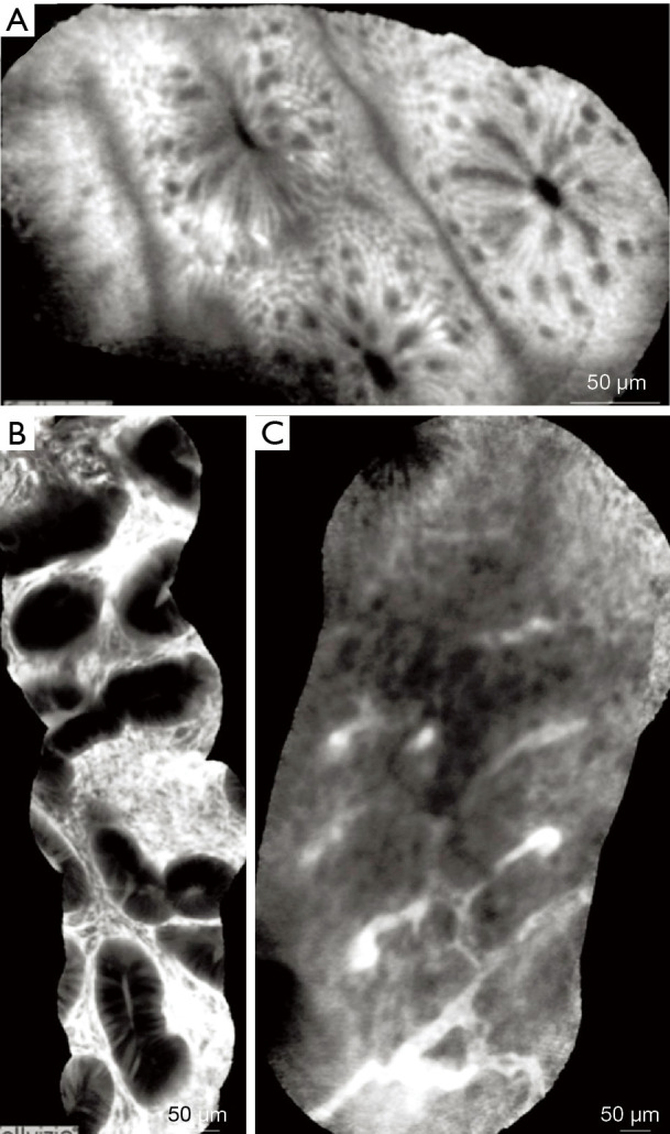
Figure 11.

CLE images of colonic mucosa. (A) Normal colonic mucosa-round shaped crypts, dark goblet cells, narrow and regular blood vessels surrounding the crypts. (B) Adenomatous polyp-irregular or villiform structures and a dark, irregularly thickened epithelium with a decreased number of goblet cells. (C) Adenocarcinoma-disorganized mucosa, lack of structure, elongated crypts, irregularly thickened epithelium, dilated and distorted blood vessels. Adapted from De Palma GD, et al., World J Gastroenterol, 2009. CLE, confocal laser endomicroscopy.
