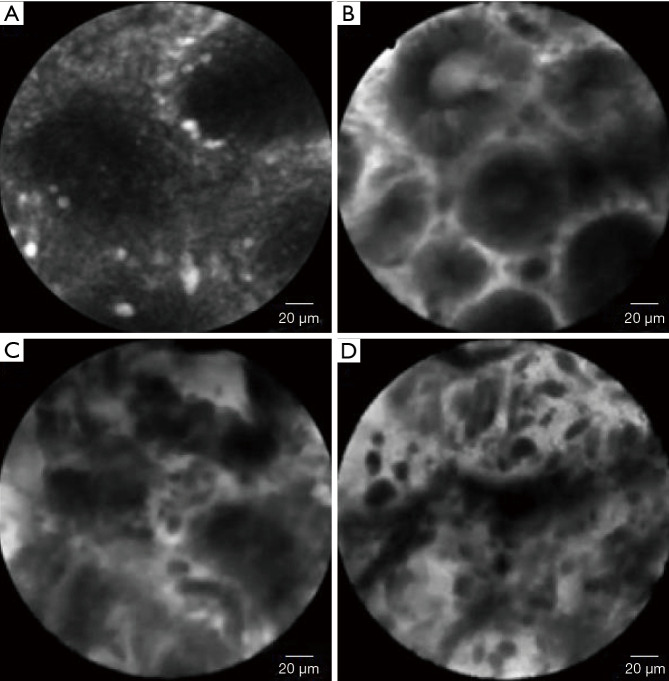
Figure 6.
The pCLE appearance of normal and pathological gastric mucosa. (A) Normal cardiac mucosa-regular pits with wide openings. (B) Dysplastic gastric-dark epithelium with irregular and varying thickness is observed. (C) Differentiated adenocarcinoma-disorganized epithelium with dark and irregular glands. (D) Undifferentiated adenocarcinoma-dark and irregular cells with no identifiable glandular structures are observed. Adapted from Kin MY, et al., World J Gastroenterol, 2016. pCLE, probe-based confocal laser endomicroscopy.

