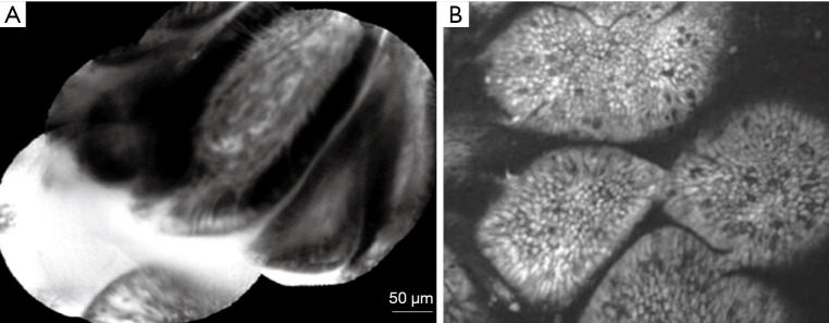
Figure 8.
The CLE appearance of normal and pathological small bowel mucosa. (A) Normal small bowel mucosa-normal epithelium border with regular capillary pattern. (B) Confocal image of celiac disease-Marsh type 3b. Magnification: 1,000×. Adapted from De Palma, GD, World J Gastroenterol, 2009; Venkatesh K, World J Gastroenterol, 2009. CLE, confocal laser endomicroscopy.

