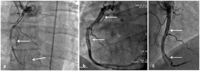
Figure 5.
Repeated coronary angiography: (a) defect of contrasting of the RCA with stenosis by 90% (upper white arrow), occlusion of the distal segment (lower white arrow); (b) and (c) linear dissections of the RCA and the initial segment of the posterior lateral branch of the RCA (indicated by arrows).

