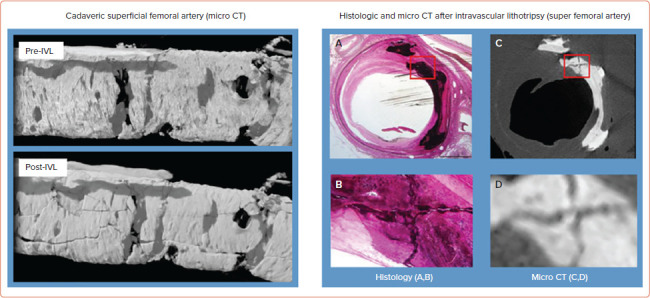
Figure 4: Calcic Microfractures Induced by Intravascular Lithotripsy in Micro CT and Histology.

Left panel: Representative micro CT images before and after intravascular lithotripsy (IVL) treatment. Abluminal view of the left distal femoral artery demonstrating predominantly medial calcification. (A) Before (A) and after (B) IVL. Circumferential, transverse, and longitudinal calcium fractures were observed following IVL treatment. Right panel: Histological and micro CT imaging after IVL treatment. Cross-sectional histological Exakt ground section (A) matched with the micro CT cross-sectional image (C). Both sections show cross-shaped cracks highlighted by red boxed areas, which are shown at high-power magnification (B,D). Source: Kereiakes et al. 2021.[8] Adapted with permission from Elsevier.
