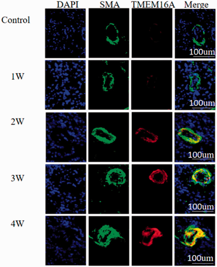
Fig. 3.
Localization of TMEM16A in the arterioles of lung tissues from MCT-treated rats. Lung tissues were isolated from rats treated with MCT for 0 (control), 1 (1W), 2 (2W), 3 (3W), and 4 weeks (4W). Nuclei were counterstained with DAPI (blue). SMA (green) indicated the location of vascular smooth muscle cells. TMEM16A was shown by red fluorescence. “Merge” represents merged SMA and TMEM16A signals. DAPI: diamidino-phenyl indole; SMA: smooth muscle α-actin; TMEM16A: transmembrane protein 16A.

