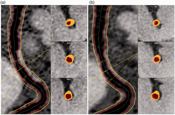
Figure 2.
Magnetic resonance vessel wall imaging (MR-VWI) was measured using a custom-designed intracranial vessel analysis software package. In the baseline examination (a), contiguous cross-sectional slices from causative lesions were reconstructed with a semi-automatic centerline tracking functionality, and vessel wall was segmented in each slice with a deep learning-based algorithm. The images from the follow-up examination (b) were spatially registered to those from the baseline, and the same centerline path and slice range involving the original causative lesion were applied to generate location-matched contiguous cross-sectional slices.

