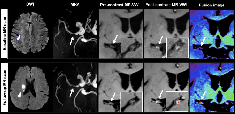
Figure 5.
A 61-year-old male patient received baseline magnetic resonance imaging (MRI) 4 days after stroke and follow-up MRI one day after recurrent stroke (arrowheads) at 10 months. The causative plaque (arrows) at the right middle cerebral artery deteriorated with increases in plaque volume (16.18%), peak normalized wall index (pNWI) (6.82%), plaque-wall contrast ratio (CR) (23.78%) and plaque enhancement ratio (ER) (26.65%). Asterisks indicate the lumen.

