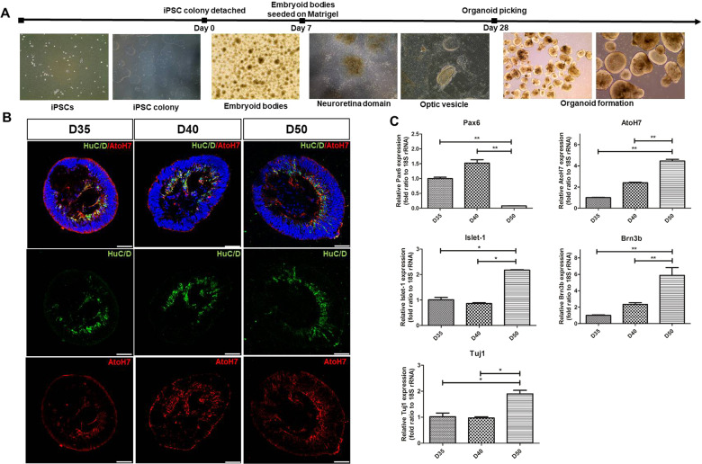
Fig. 1. Formation of hiPSC-derived ROs and RGC development at an early stage.
A Main steps of hiPSC-derived RO development in vitro: hiPSC colony, EB formation, neuroretinal domain, and ROs. B Immunofluorescence staining of HuC/D (green) and AtoH7 (red) of ROs at distinct differentiated time points. The expression of HuC/D and AtoH7 was gradually increased in the innermost layers of ROs from day 35 to day 60. C Quantitative RT-PCR results of PAX6, AtoH7, Islet-1, Brn3b, and Tuj1 levels of ROs at indicated time points. Data are expressed by mean ± S.E.M. *p < 0.05, **p < 0.01 by the Kruskal–Wallis test with post hoc analysis. Scale bar, 50 μm.

