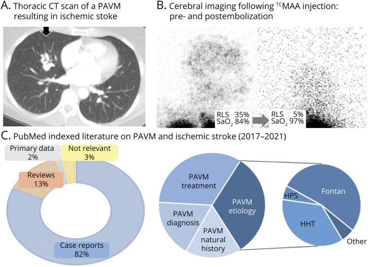
Figure 1. Pulmonary Arteriovenous Malformations and Right to Left Shunts.
(A) Thoracic CT scan of a pulmonary arteriovenous malformation (PAVM) in a patient admitted with acute ischemic stroke. (B) Diagnosis and quantification of pathologic right-to-left shunt (RLS) on right lateral head projections following injection of 99mTc-labeled albumin macroaggregates in a patient pre and post PAVM embolization, resulting in a visible reduction of shunt size and corresponding improvement in oxygen saturation (SaO2) levels.2,3 Note the intense activity in the lung apices is as expected, but that in the postembolization image, although the gain has been turned up, trivial cerebral activity is still visible. (C) PubMed-indexed English language literature (1 January 2017–April 3, 2021) on “pulmonary arteriovenous malformation” and “stroke” depicting a publication bias favoring case reports, especially those describing rare associations; for example, Fontan circulation and hepatopulmonary syndrome (HPS), rather than hereditary hemorrhagic telangiectasia (HHT), which is responsible for PAVMs in >70% of cases.4

