
Extended Data Fig. 9 |. Ordered C terminus in CaSR and structural comparison between CaSR and mGlu2.
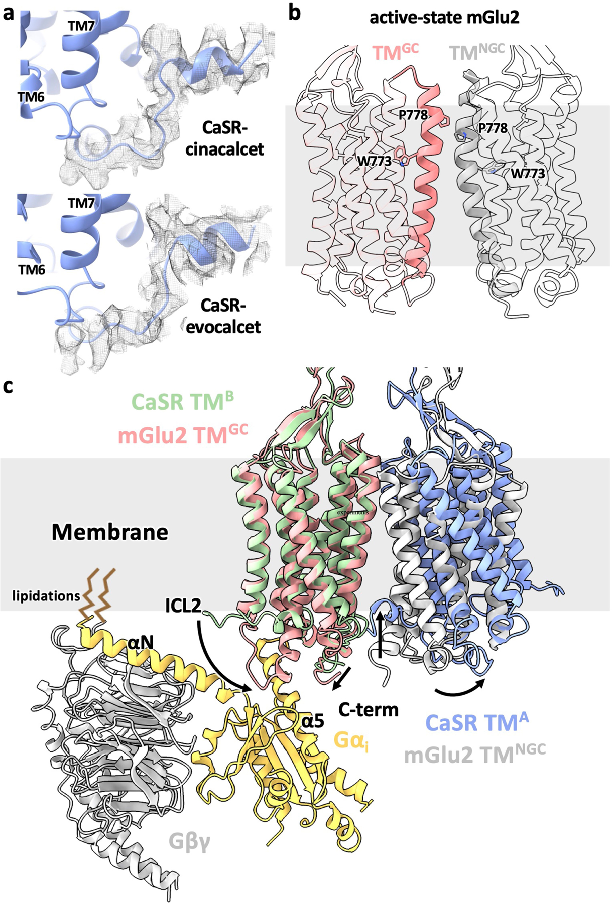
a, The ordered C terminus from the 7TM with a bent PAM (7TMB) shown in cryo-EM densities with unsharpened maps. b, Asymmetric 7TMs in the active-state mGlu2 alone structure (accompanying manuscript). TM6 are shown as solid cartoon with representative residues shown as sticks to highlight the asymmetry. c, Superposition of the G protein-coupling 7TM (7TMGC) from mGlu2-Gi complex structure (accompanying manuscript) onto the active-state CaSR 7TM with a straight PAM (7TMA) showing that the G protein would fit well on the membrane plane and the tilt of CaSR 7TMB leads to the sequestration of its C terminus in the membrane. The comparison between mGlu 7TMGC and CaSR 7TMA illustrates that the receptor likely would couple to G proteins through downward extensions of both ICL2 and C terminus.