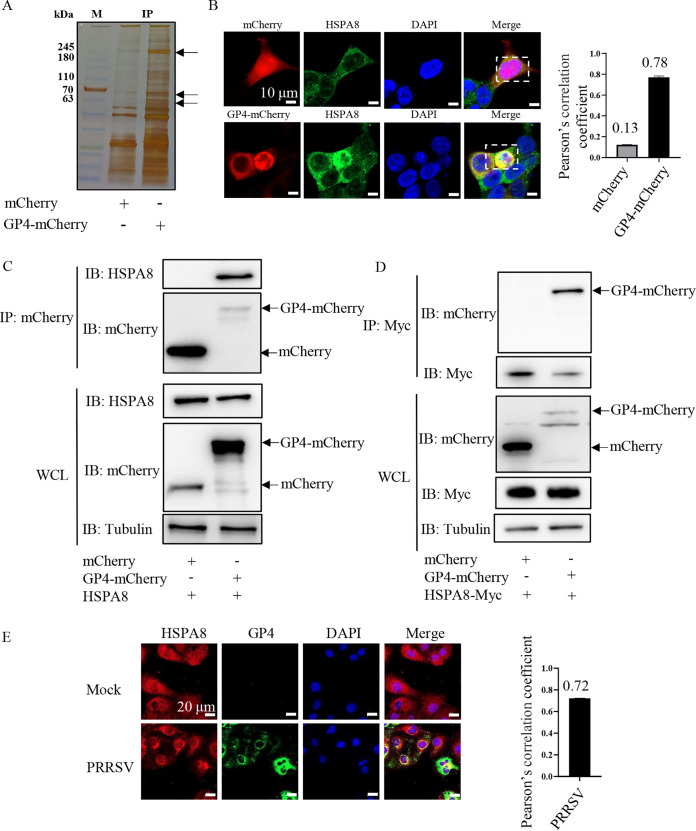
FIG 1.
Identification of HSPA8 interacting with PRRSV GP4. (A) Silver staining of the associated proteins with GP4-mCherry. The HEK-293T cells were transfected with the plasmid expressing mCherry or GP4-mCherry. The proteins were immunoprecipitated in WCLs using anti-mCherry antibody, then separated by 12% SDS-PAGE and stained with silver. The arrows indicated different immunoprecipitated protein bands in the GP4-mCherry-expressed cells from in the mCherry-expressed ones. Lane M, protein marker. (B) GP4-mCherry co-localized with endogenous HSPA8. HEK-293T cells were transfected with the plasmid expressing GP4-mCherry (red) or mCherry (red) for 24 h, and stained with anti-HSPA8 pAbs (catalog no. 10654-1-AP; green). The cell nuclei were stained with DAPI (blue). The co-localization was assessed by determination of Pearson’s correlation coefficient. Scale bars, 10 μm. (C) GP4-mCherry interacted with endogenous HSPA8. HEK-293T cells were transfected with the plasmids expressing mCherry and GP4-mCherry, respectively. MCherry or GP4-mCherry was immunoprecipitated from WCLs by anti-mCherry antibody and their immunoprecipitated proteins were immunoblotted with anti-HSPA8 pAbs (catalog no. 10654-1-AP) and anti-mCherry pAbs. (D) GP4-mCherry interacted with exogenous HSPA8. HEK-293T cells were co-transfected with the plasmids expressing HSPA8-Myc, and mCherry or GP4-mCherry, respectively. HSPA8-Myc immunoprecipitated proteins were immunoblotted with anti-mCherry pAbs and anti-Myc MAb. (E) The endogenous co-localization between HSPA8 and PRRSV GP4 in the infected MARC-145 cells. MARC-145 cells were infected with PRRSV at 0.1 MOI for 24 h, and stained with anti-HSPA8 pAbs (catalog no. 10654-1-AP; green) and anti-GP4 pAbs (green). Nuclei were stained with DAPI (blue). The co-localization was assessed by determination of Pearson’s correlation coefficient. Scale bars, 20 μm.

