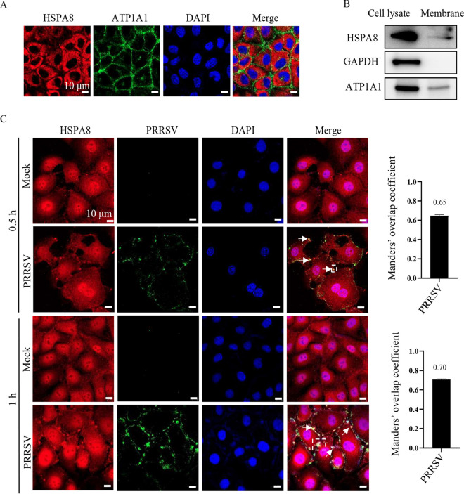
FIG 4.
HSPA8 co-localizes with PRRSV during attachment and internalization in MARC-145 cells. (A, B) HSPA8 was expressed both on the surface and in the cytoplasm of MARC-145 cells. (A) MARC-145 cells were fixed with PFA, and stained with anti-HSPA8 MAb (catalog no. 66442-1-AP; red) and anti-ATP1A1 pAbs (green), respectively. The cell nuclei were stained with DAPI. Images were acquired on the Zeiss confocal microscope. Scale bars, 10 μm. (B) MARC-145 cell membranes were extracted. WCLs and membrane extracts were subjected to IB using anti-HSPA8 pAbs (catalog no. 10654-1-AP), anti-ATP1A1 pAbs, and anti-GAPDH pAbs, respectively. (C) PRRSV co-localized with HSPA8 in MARC-145 cells during attachment and internalization. MARC-145 cells were infected with PRRSV (10 MOI) at 37°C for 0.5 and 1 h. The cells were washed with PBS, fixed with 4% PFA, permeabilized with 0.1% Triton X-100, and stained with anti-HSPA8 pAbs (catalog no. 10654-1-AP; red) and anti-PRRSV GP5 MAb (green). The cell nuclei were stained with DAPI (blue). Images were acquired on the confocal microscope with the same confocal microscope settings. The white arrows indicated the clusters of HSPA8 with PRRSV. The Manders’ overlap coefficient in white dashed line box was analyzed. The mock-infected cells were used as control. Scale bars, 10 μm.

