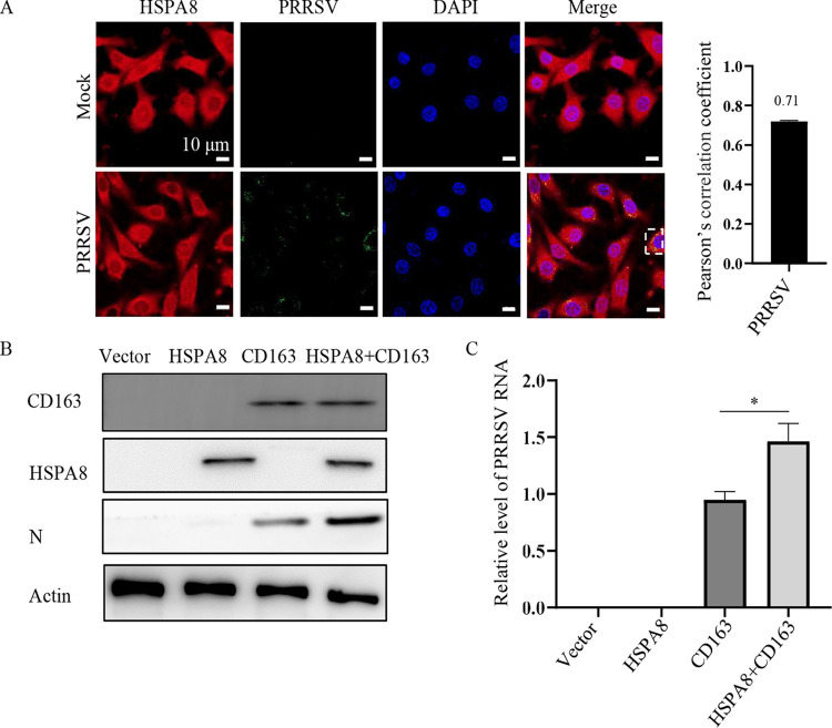
FIG 9.
HSPA8 contributes to PRRSV infection along with CD163. (A) HSPA8 co-localized with internalized PRRSV in BHK-21 cells. The cells were inoculated with PRRSV (10 MOI) at 37°C for 0.5 h, and fixed with 4% PFA, permeabilized with 0.1% Triton X-100 and stained with anti-HSPA8 pAbs (catalog no. 10654-1-AP; red) and anti-PRRSV GP5 MAb (green). The cell nuclei were stained with DAPI (blue). Images were acquired on the Zeiss confocal microscope. The co-localization was assessed by determination of Pearson’s correlation coefficient. Scale bars, 10 µm. (B-C) HSPA8 contributed to PRRSV infection. BHK-21 cells were transfected with HSPA8-Myc and/or CD163 for 36 h and then infected with PRRSV (1 MOI) for 24 h. The cells were subjected to IB using anti-Myc MAb, anti-CD163 pAbs, anti-actin MAb, and anti-PRRSV N MAb (B). PRRSV RNA abundance was determined by RT-qPCR (C). Data represent means ± SD from three independent experiments. *, P < 0.05.

