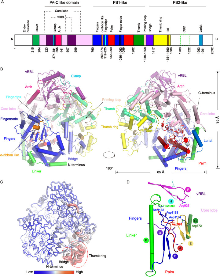
FIG 3.
Overall structure of the RVFV L protein and conserved motifs. (A) Schematic representation of the monomeric RVFV L protein domain structure. The white areas are missing in the determined structure but present in the purified protein. (B) Cartoon representation of the RVFV L protein. The structure is colored by domains using the same color code as in (A). 310 helices are colored in gray. (C) The RVFV L protein B-factor map. A larger radius and red color represent high B-factor values and a smaller radius and blue color represent low B-factor values. (D) The arrangement of the conserved RdRp motifs in the RVFV active site colored in red, green, blue, purpleblue, yelloworange, magenta, forest, and teal for motifs A–H, respectively.

